ERCP Treatment in Nashik

With the scope in this position, the major duodenal papilla is identified and inspected for abnormalities. This structure is a protrusion of the hepatopancreatic ampulla (also known as the ampulla of Vater) into the duodenal lumen. The ampulla is the convergence point of the ventral pancreatic duct and common bile duct (CBD) and thus acts as a conduit for drainage of bile and pancreatic secretions into the duodenum.
The minor duodenal papilla is also located in the second portion of the duodenum and serves as the access point for the dorsal pancreatic duct. Evaluation of the dorsal pancreatic duct with ERCP is rarely performed; indications are discussed below.
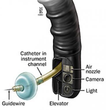
After the papilla has been examined with the side-viewing endoscope, selective cannulation of either the CBD or the ventral pancreatic duct is performed. Once the chosen duct is cannulated, either a cholangiogram (CBD) or a pancreatogram (pancreatic duct) is obtained fluoroscopically after injection of radiopaque contrast material into the duct. ERCP is now primarily a therapeutic procedure; thus, abnormalities that are visualized fluoroscopically can typically be addressed by means of specialized accessories passed through the working channel of the endoscope.
Because ERCP is an advanced technique, it is associated with a higher frequency of serious complications than other endoscopic procedures are. Accordingly, specialized training and equipment are required, and the procedure should be reserved for appropriate indications.
A review by Freeman et al, using data from 2004, estimated that about 500,000 procedures were performed annually in the United States. [1] However, because of a decrease in diagnostic ERCP with the advent of endoscopic ultrasonography (EUS) and magnetic resonance cholangiopancreatography (MRCP), this number is likely decreasing.
Make An Appointment
Book your appointment with our consultants.
Get in Touch
91 9923825259 / +91 9822011684
0253 2574070 / 0253 2572421
By e-Mail:
nitinborse@hotmail.com
